What Is Cardiac Imaging, Testing & Diagnostics?
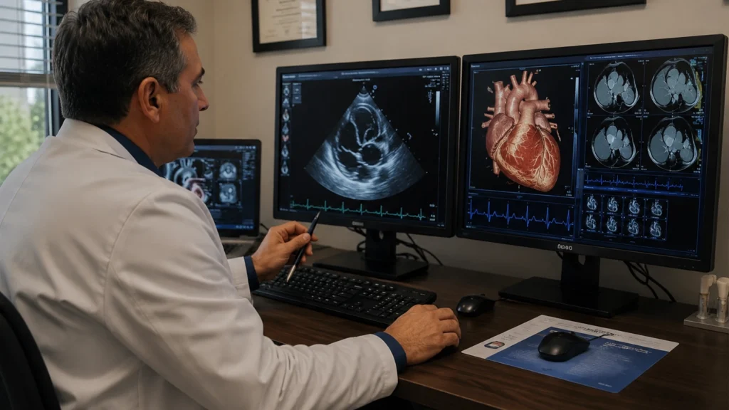
Cardiac imaging and diagnostic testing is the collection of tools cardiologists use to evaluate the structure and function of your heart and blood vessels without surgery. No single test answers every question; that’s why Florida Heart & Vascular Institute offers multiple modalities at our Ocala, FL location and beyond, selecting the right test based on symptoms, history, and clinical suspicion.
Some tests produce images of your heart’s anatomy, showing chamber size, valve function, and muscle movement. Others record electrical activity, track blood flow under stress, or monitor rhythm over hours or years. At FHVI’s Ocala offices, diagnostic testing and result interpretation happen under one roof, shortening the time between your symptom and your treatment plan.
Echocardiography
A heart ultrasound that evaluates chamber size, pumping function (ejection fraction), valve movement, and blood flow. A standard transthoracic echo (TTE) places a probe on the chest wall. A transesophageal echo (TEE) passes a probe through the oesophagus for sharper images — used before cardioversion, Watchman implantation, or when TTE results are unclear.
EKG and Rhythm Testing
An EKG records your heart’s electrical activity in about 10 minutes. When symptoms like palpitations, dizziness, or fainting occur intermittently and don’t appear on a short office EKG, a Holter monitor (24-48 hours), event recorder (2-4 weeks), or implantable LOOP recorder (up to 3 years) captures the rhythm at the right moment.
Stress Testing
Exercise or pharmacological stress testing shows how the heart performs under strain. A standard treadmill test records the EKG during exercise. A nuclear stress test or stress echocardiogram adds imaging to assess blood flow and heart muscle function — detecting coronary artery disease that a plain EKG misses.
Cardiac CT and Coronary Calcium Scoring
A coronary calcium score scan is a fast, low-radiation CT that measures calcified plaque in the coronary arteries — useful for risk stratification in asymptomatic patients. A CT coronary angiogram (CTCA) provides a non-invasive view of the coronary artery lumen, an alternative to diagnostic cardiac catheterization in low-to-intermediate-risk patients.
Cardiac MRI
MRI provides detailed images of heart muscle texture, scarring, and inflammation without radiation. Gold standard for myocarditis, cardiac sarcoidosis, and hypertrophic cardiomyopathy. Takes 45 to 75 minutes.
Nuclear Imaging and Vascular Studies
Nuclear cardiac imaging uses a small radioactive tracer to assess heart muscle blood flow at rest and during stress. Vascular ultrasound — carotid duplex, arterial and venous duplex, ABI testing, and AAA screening — evaluates the arteries and veins beyond the heart when peripheral or cerebrovascular disease is suspected.
When You May Need Cardiac Imaging or Diagnostic Testing
Cardiac testing doesn’t only happen when your doctor suspects a serious problem. It’s used to rule out disease, establish a baseline, track a known condition, and screen high-risk patients before symptoms develop.
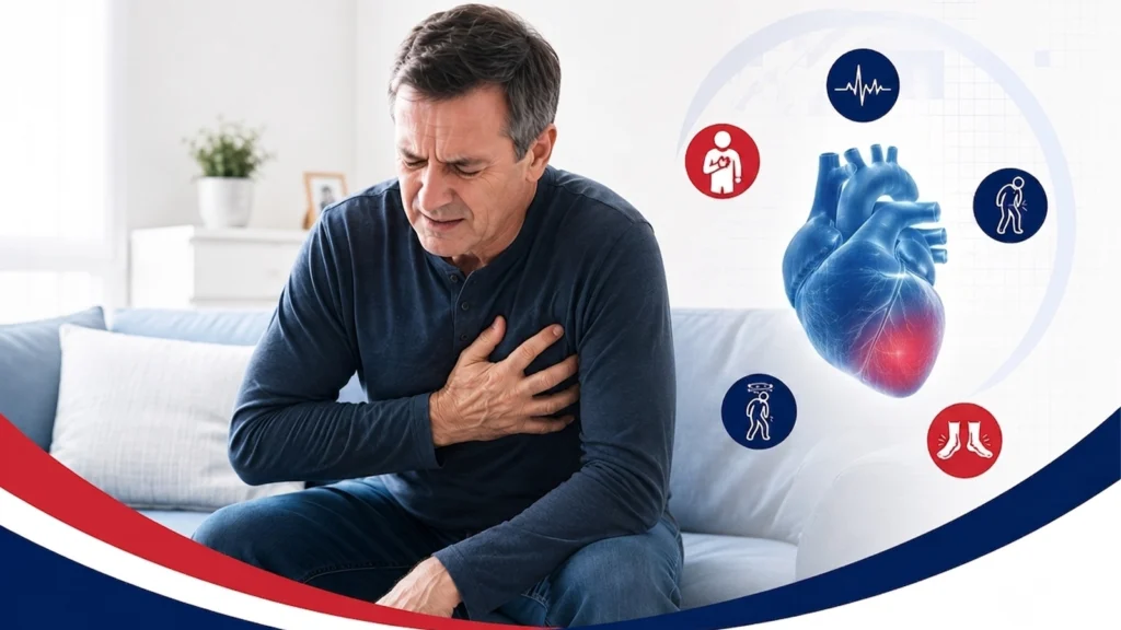
Get tested urgently, if you have:
- Chest pain or pressure with exertion that eases with rest — classic coronary artery disease warning sign
- New or worsening shortness of breath at rest or with minimal activity
- Palpitations, racing heart, or skipped beats — new, frequent, or lasting over 30 seconds
- Swollen ankles or legs combined with breathlessness
- An abnormal EKG or murmur found by your primary care doctor
Get screened proactively, if you have:
- High blood pressure not yet evaluated from a cardiac standpoint
- Type 2 diabetes — doubles cardiovascular risk
- High cholesterol with multiple other risk factors
- Family history of heart attack or sudden cardiac death before age 55
- Prior heart attack, stent, or bypass surgery
- Atrial fibrillation (AFib) requiring monitoring
Emergency Warning
Chest pain with sweating, nausea, severe shortness of breath, fainting, or pain spreading to the arm, jaw, neck, or back may be a heart attack. Call 911 immediately. Do not drive yourself.
Risks and Benefits of Cardiac Imaging and Diagnostic Testing
Most cardiac imaging tests are non-invasive, carrying no procedural risk at all. Understanding each test helps patients make confident decisions.
Benefits
- Earlier diagnosis — catch problems before irreversible damage
- Accurate treatment decisions matched to the specific finding
- Anxiety reduction — a clear result ends months of uncertainty
- Disease monitoring — track progression and treatment response
- Outpatient convenience — no hospital admission needed
Risks
- Echo, EKG, Holter, ABI, vascular ultrasound: No risk
- Stress test: very low risk; staff monitor continuously
- Nuclear stress test: radiation dose comparable to chest CT
- Cardiac CT: low-dose radiation; contrast reviewed for kidney patients
- Cardiac MRI: no radiation; gadolinium contrast reviewed for kidney function
- TEE: small risk of throat irritation; sedation required
Get the cardiac answers you've been waiting for
FHVI’s board-certified cardiologists perform and interpret the full range of cardiac imaging at our Ocala flagship and 15 Florida locations. Same-week appointments are available for urgent symptoms.
Cardiac Imaging, Testing and Diagnostic Procedures at FHVI
Every test below is available at FHVI without a referral to an outside imaging centre. Your cardiologist orders the study, our team performs it, and the same physician reads the result — so you get one point of contact from test to treatment plan. Most procedures need little to no preparation, take under an hour, and let you go home the same day.
Echocardiogram (Echo)
Uses ultrasound to evaluate your heart's chambers, valves, pumping strength, and ejection fraction. Available as transthoracic (TTE) or transesophageal (TEE) studies depending on your condition.
Electrocardiogram (EKG)
Records your heart's electrical activity to detect abnormal rhythms, previous heart attacks, and conduction disorders.
Exercise Stress Test (Treadmill)
Measures how your heart performs during exercise to evaluate blood flow, rhythm changes, and exercise tolerance.
Nuclear Stress Test
Uses a small amount of radioactive tracer to compare blood flow to your heart at rest and during stress, helping identify blocked arteries.
Stress Echocardiogram
Combines ultrasound imaging with exercise or medication induced stress to evaluate heart muscle function and blood flow.
Holter Monitor
Continuously records your heart rhythm during normal daily activities to detect intermittent rhythm abnormalities.
Event Monitor
Records your heart rhythm during symptoms or automatically when an abnormal rhythm occurs, helping diagnose infrequent heart rhythm problems.
Implantable Loop Recorder
A small device placed under the skin continuously monitors your heart rhythm for up to three years to detect unexplained fainting or rare arrhythmias.
Coronary Calcium Score
A specialized CT scan that measures calcium buildup in your coronary arteries to assess your future risk of heart disease.
CT Coronary Angiography (Cardiac CTA)
Uses contrast enhanced CT imaging to produce detailed pictures of your coronary arteries without requiring cardiac catheterization.
Cardiac MRI
Provides detailed images of your heart muscle, valves, and surrounding structures without radiation, helping diagnose many complex heart conditions.
Carotid Duplex Ultrasound & Vascular Studies
Uses ultrasound to evaluate blood flow in the carotid arteries, leg arteries and veins, or abdominal aorta to detect narrowing, blockages, aneurysms, or blood clots.
Cardiac Imaging & Diagnostics Near Me — FHVI Locations
Florida Heart & Vascular Institute’s flagship location is in Ocala, Florida, with 14 additional locations across North Central, Central, and Southwest Florida. Patients searching for ‘cardiac imaging Ocala Florida’ or ‘heart testing near me’ can access full diagnostic testing close to home.
Why Patients Choose Florida Heart & Vascular Institute
One Practice. Full Diagnostic Spectrum
Complete cardiac imaging under one roof with no outside referrals or unnecessary delays.
Board Certified Cardiologists
Your imaging studies are interpreted by experienced board certified cardiovascular specialists.
15 Convenient Locations in Florida
High quality heart care close to home throughout Central and North Florida.
Fast Results. Faster Treatment
Quick turnaround on reports helps your physician begin treatment sooner.
Advanced Technology You Can Trust
State of the art imaging equipment delivers accurate and reliable diagnostic results.
Patient Centered Care Florida
Every patient receives compassionate, personalized care from diagnosis through treatment.
Frequently Asked Questions
Cardiac imaging is the broad term for all tests used to see inside and around the heart without surgery. It includes ultrasound (echocardiogram), nuclear imaging, CT, MRI, and EKG. Each test answers a different clinical question, and your FHVI cardiologist in Ocala selects the most appropriate test based on your symptoms.
An echocardiogram uses sound waves to create real-time images of the heart. A gel is applied to your chest and a probe is moved across the surface. It takes 30 to 45 minutes and causes no discomfort for most patients.
An EKG records the electrical activity of the heart. It detects atrial fibrillation, evidence of a prior heart attack, heart block, and irregular rhythms. Takes about 10 minutes with no needles.
A standard exercise stress test records your EKG while you walk on a treadmill. A nuclear stress test adds radioactive tracer imaging showing blood flow to the heart muscle, making it more sensitive for detecting blocked arteries.
An EKG takes 10 minutes. An echocardiogram takes 30-45 minutes. A stress test takes 45-60 minutes. A nuclear stress test takes 3-4 hours. A cardiac MRI takes 45-75 minutes.
A fast, low-radiation CT scan counting calcified plaque in coronary arteries. Used for risk stratification in patients with borderline cholesterol or family history.
Yes. FHVI accepts Medicare, Medicaid, and most major insurance plans at all locations including Ocala. Call 352-619-2550 to verify coverage.
FHVI accepts referrals from primary care physicians. You may also schedule a cardiology consultation directly at our Ocala location.
FHVI’s flagship location is at 1609 SW 17th Street, Suite 100, Ocala, FL 34471. A second Ocala office is at 7535 SW 62nd Court, and a third at 1805 SE Lake Weir Avenue.
A portable EKG recorder worn continuously for 24-48 hours. Ordered when symptoms like palpitations don’t appear during a standard office EKG.
Provides the most detailed images of heart muscle structure and scarring. Used when echocardiogram results are unclear or a specific diagnosis like myocarditis is suspected.
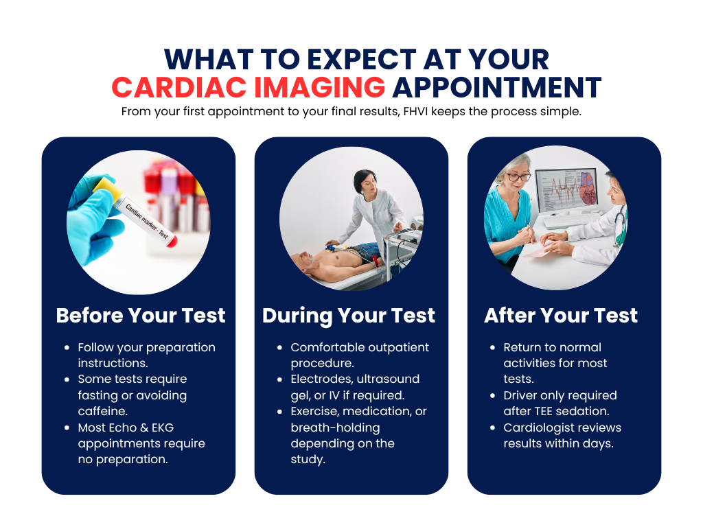
Preparation varies by test. EKGs and echocardiograms need no preparation. Stress tests require fasting 3 hours. Nuclear stress tests require no caffeine for 4-24 hours. Your FHVI scheduling team gives specific instructions when booking.
That’s a question your FHVI cardiologist answers after reviewing your symptoms, history, and risk factors. There’s no single ‘best’ test — the right one is matched to your clinical question.
Cardiac CT is faster and uses X-rays, best for coronary artery imaging. Cardiac MRI uses magnetic fields, no radiation, superior for muscle tissue evaluation.

