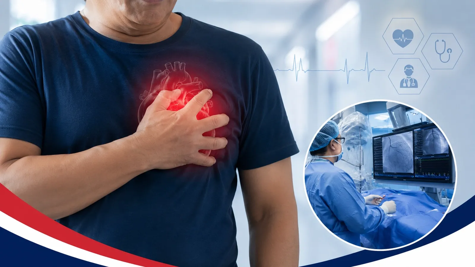
What Is Cardiac Imaging & Diagnostic Testing?

Cardiac imaging and diagnostic testing help cardiologists evaluate the structure, function, blood flow, and electrical activity of the heart and blood vessels.

Different tests answer different clinical questions. Depending on your symptoms, medical history, and cardiovascular risk, your care team may recommend imaging, diagnostic testing, vascular studies, laboratory testing, or additional cardiovascular evaluation.

Cardiac imaging and diagnostic testing at Florida Heart & Vascular Institute

At FHVI, diagnostic testing is used to help identify cardiovascular conditions, monitor known disease, and support individualized treatment planning.

Echocardiography

Echocardiography uses ultrasound to create images of the heart and evaluate chamber size, pumping function, valve movement, and blood flow. A transthoracic echocardiogram (TTE) is performed using an ultrasound probe on the chest. In selected patients, a transesophageal echocardiogram (TEE) may provide more detailed images when additional evaluation is needed.

Electrocardiogram (EKG)

An electrocardiogram records the electrical activity of the heart and may help identify rhythm abnormalities, conduction problems, or other changes that require additional cardiovascular evaluation. If longer-term rhythm monitoring is needed, your care team may recommend additional cardiac monitoring based on your symptoms.

Stress Testing

Stress testing evaluates how the heart responds to physical exercise or medication-induced stress. Depending on the clinical question, testing may include electrocardiographic monitoring or cardiac imaging. FHVI provides dedicated stress testing services for selected patients based on symptoms, cardiovascular risk, and prior test results.

Cardiac CT Imaging

Cardiac CT can provide detailed images of the heart and coronary arteries in selected patients. The type of imaging recommended depends on symptoms, cardiovascular risk, previous testing, kidney function, and the clinical question being evaluated.

Cardiac MRI

Cardiac MRI can provide detailed images of heart muscle, structure, and tissue characteristics without ionizing radiation. It may be recommended in selected patients when additional information is needed to evaluate certain cardiovascular conditions.

PET Cardiac Imaging

PET cardiac imaging uses a small amount of radioactive tracer together with specialized imaging technology to evaluate blood flow to the heart muscle and support the assessment of selected cardiovascular conditions. Your cardiologist will determine whether PET cardiac imaging is appropriate based on your symptoms, medical history, previous testing, and overall cardiovascular risk.

Vascular Doppler Ultrasound

Vascular Doppler ultrasound is a noninvasive test that uses sound waves to evaluate blood flow through arteries and veins. It may help identify narrowing, blockages, circulation problems, or other vascular abnormalities that require further evaluation.

In-House Laboratory Testing

Laboratory testing can provide important information about cardiovascular risk factors and overall health. Depending on your care plan, testing may help evaluate cholesterol, blood sugar, kidney function, blood counts, or other measurements relevant to cardiovascular diagnosis and treatment.

Availability and specific testing may vary by location and clinical need.

When You May Need Cardiac Imaging or Diagnostic Testing

Cardiac imaging and diagnostic testing may be recommended when symptoms, risk factors, physical examination findings, or previous test results suggest that more information about your heart or circulation is needed. Testing may also be used to monitor a known cardiovascular condition or evaluate changes over time.

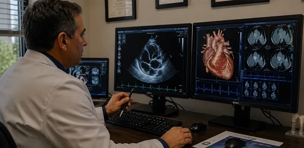
Patient discussing heart symptoms and diagnostic testing with a cardiologist

Consider Diagnostic Evaluation If You Have:

  • Chest discomfort or pressure
  • New or worsening shortness of breath
  • Unexplained fatigue or reduced exercise tolerance
  • Dizziness, fainting, or recurrent palpitations
  • Leg swelling or other signs of fluid retention
  • An abnormal EKG, heart murmur, or other examination finding
  • New cardiovascular symptoms with known heart disease

Testing May Also Be Recommended If You Have:

  • High blood pressure
  • High cholesterol
  • Diabetes or other cardiovascular risk factors
  • A family history of cardiovascular disease
  • A previous heart attack, coronary stent, or bypass surgery
  • Known heart valve disease
  • A known cardiovascular condition requiring follow-up

Emergency Warning

Call 911 immediately for severe or persistent chest pain, severe shortness of breath, fainting, stroke-like symptoms, or other symptoms that may indicate a medical emergency.

Risks and Benefits of Cardiac Imaging & Diagnostic Testing

Cardiac imaging and diagnostic testing can provide important information about heart structure, function, circulation, and cardiovascular risk. The potential benefits and risks vary depending on the test recommended and each patient’s individual health needs. Before testing, your care team will explain what to expect, whether preparation is needed, and any risks or considerations specific to your test.

Benefits

Risks

Need More Clarity About Your Heart Health?

Cardiac imaging and diagnostic testing can help your care team better understand your symptoms, heart function, circulation, and cardiovascular risk. FHVI can help determine which test or evaluation may be appropriate for your individual needs.

Cardiac Imaging & Diagnostics

Cardiac Imaging, Testing & Diagnostic Services

FHVI provides cardiovascular imaging and diagnostic services to help evaluate heart function, blood flow, vascular health, and cardiovascular risk. Testing is selected based on each patient’s symptoms, medical history, prior results, and clinical needs.

Core Cardiac Imaging & Diagnostic Services

These services help evaluate heart structure, function, circulation, vascular health, and cardiovascular risk to support diagnosis and treatment planning.
PET cardiac imaging

PET Cardiac Imaging

Advanced Imaging

PET cardiac imaging uses a small amount of radioactive tracer and specialized imaging technology to evaluate blood flow to the heart muscle and support assessment of selected cardiovascular conditions.

Blood Flow Evaluation
Cardiac Imaging
Diagnostic Support
Echocardiography

Echocardiography

Heart Ultrasound

Echocardiography uses ultrasound to create images of the heart and evaluate chamber size, pumping function, valve movement, and blood flow. Depending on clinical needs, testing may include transthoracic or transesophageal echocardiography.

Heart Structure
Valve Evaluation
No Ionizing Radiation
Electrocardiogram EKG

Electrocardiogram (EKG)

Electrical Testing
An electrocardiogram records the electrical activity of the heart and may help identify rhythm abnormalities, conduction problems, or other changes that require additional cardiovascular evaluation.

Heart Rhythm
Noninvasive
Diagnostic Testing
Cardiovascular diagnostic testing

Cardiovascular Diagnostic Testing

Diagnostic Evaluation

Cardiovascular diagnostic testing may include selected studies used to evaluate heart function, circulation, symptoms, and cardiovascular risk. Testing is chosen based on the clinical question and the patient’s individual care needs.

Heart Function
Clinical Evaluation
Care Planning
Vascular Doppler ultrasound

Vascular Doppler Ultrasound

Vascular Imaging
Vascular Doppler ultrasound is a noninvasive test that uses sound waves to evaluate blood flow through arteries and veins. It may help identify narrowing, blockages, or other circulation-related abnormalities.

Blood Flow Evaluation
Noninvasive
No Ionizing Radiation
In-house laboratory testing

In-House Laboratory Testing

Laboratory Testing

Laboratory testing can provide important information about cardiovascular risk factors and overall health. Depending on the care plan, testing may help evaluate cholesterol, blood sugar, kidney function, blood counts, and other clinically relevant measurements.

Risk Assessment
Blood Testing
Care Planning
Cardiac CT and coronary calcium scoring

Cardiac CT & Coronary Calcium Scoring

CT Imaging
Cardiac CT can provide detailed images of the heart and coronary arteries. Coronary calcium scoring may help assess calcified plaque burden and support cardiovascular risk evaluation in selected patients.

Coronary Arteries
Risk Assessment
CT Imaging
CT coronary angiography

CT Coronary Angiography

Coronary Imaging

CT coronary angiography uses CT imaging and contrast material to create detailed images of the coronary arteries. It may be recommended in selected patients when additional evaluation of coronary anatomy is clinically appropriate.

Coronary Anatomy
Contrast Imaging
Diagnostic Evaluation
Cardiac MRI

Cardiac MRI

Advanced Imaging

Cardiac MRI provides detailed images of heart muscle, structure, and tissue characteristics without ionizing radiation. It may be used in selected patients when additional information is needed to evaluate certain cardiovascular conditions.

Heart Structure
Tissue Evaluation
No Ionizing Radiation

Related Cardiac Testing & Monitoring

Some patients may also need stress testing or longer-term rhythm monitoring. These services are part of FHVI’s broader cardiovascular diagnostic pathway and may also be available through dedicated specialty services.
Treadmill stress testing

Treadmill Stress Testing

Stress Testing
Treadmill stress testing evaluates how the heart responds to physical activity while heart rate, rhythm, blood pressure, and symptoms are monitored.

Exercise Testing
Heart Response
Diagnostic Testing
Nuclear stress testing

Nuclear Stress Testing

Stress Imaging

Nuclear stress testing combines stress evaluation with specialized imaging to assess blood flow to the heart muscle. The test may use exercise or medication-induced stress depending on the patient’s clinical needs.

Blood Flow Imaging
Stress Evaluation
Nuclear Imaging
Stress echocardiography

Stress Echocardiography

Stress Imaging

Stress echocardiography combines ultrasound imaging with exercise or medication-induced stress to evaluate how the heart functions under increased workload.

Ultrasound Imaging
Heart Function
Stress Evaluation
Holter heart rhythm monitor

Holter Monitoring

Rhythm Monitoring

A Holter monitor continuously records the heart’s electrical activity over an extended period and may help identify rhythm abnormalities that are not captured during a standard EKG.

Heart Rhythm
Continuous Monitoring
Noninvasive
Cardiac event monitor

Event Monitoring

Rhythm Monitoring

Event monitoring records heart rhythm information over a longer period and may be useful when symptoms such as palpitations, dizziness, or irregular heartbeats occur intermittently.

Intermittent Symptoms
Heart Rhythm
Extended Monitoring
Implantable loop recorder for heart rhythm monitoring

Implantable Loop Recorder

Long-Term Monitoring

An implantable loop recorder is a small device placed beneath the skin to provide long-term heart rhythm monitoring. It may be recommended when symptoms are infrequent or difficult to capture with shorter-term monitoring.

Long-Term Monitoring
Heart Rhythm
Intermittent Symptoms
Important: Imaging, diagnostic testing, monitoring, and provider availability may vary by location and individual clinical needs. Your care team will recommend testing based on your symptoms, medical history, previous results, and overall cardiovascular health. Please contact FHVI to confirm the appropriate test and location for your care.
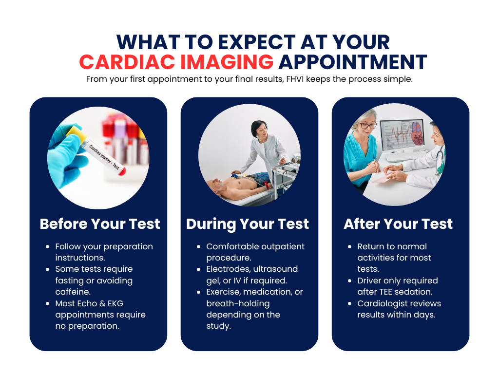
What to expect before, during, and after a cardiac imaging appointment
Meet Your Cardiac Imaging Team

Experienced Providers for Cardiovascular Imaging & Diagnostic Care

FHVI’s physicians and advanced practice providers use cardiovascular imaging and diagnostic testing to evaluate symptoms, interpret clinical findings, and guide individualized care plans.

Dr. Asad Qamar, MD

Interventional Cardiologist

David Edmund Ruggieri, MD

David Edmund Ruggieri, MD

Cardiologist

Jihad A. Mustapha, MD

Interventional Cardiologist

Syed Imran Ali, MD

Interventional Cardiologist

Cardiac Imaging Across Florida

Find an FHVI Cardiac Imaging & Diagnostic Location Near You

FHVI provides cardiovascular imaging and diagnostic testing at select locations across Florida. Test and provider availability may vary by location, so our scheduling team can help connect you with the appropriate location for your diagnostic needs.

Request Your Appointment

Complete the form below with your preferred cardiologist, location, and reason for visit. An FHVI team member will contact you to confirm availability and next steps.


    0/1000

    By submitting this form, you agree to our Privacy Policy. This form is for non-urgent inquiries only. For medical emergencies, please call 911.

    Why Choose FHVI

    Trusted Cardiovascular Care Since 1997

    Since 1997, Florida Heart & Vascular Institute has provided physician-led cardiovascular care with a focus on clinical expertise, advanced technology, clear communication, and individualized treatment. Our team works to make every step of the patient journey—from evaluation and diagnosis to treatment and follow-up—more coordinated and understandable.

    Serving Patients Since 1997

    Decades of cardiovascular care experience have shaped a patient-first approach focused on quality, continuity, and trust.

    Physician-Led Cardiovascular Care

    Our physicians stay closely involved in evaluation, treatment planning, and follow-up.

    Advanced Diagnostic Technology

    Modern cardiac and vascular testing supports accurate evaluation and informed care decisions.

    Personalized Treatment Plans

    Care recommendations are based on each patient’s symptoms, medical history, diagnostic findings, and individual needs.

    Minimally Invasive Care Options

    When appropriate, FHVI offers catheter-based and other minimally invasive treatment options for selected cardiovascular conditions.

    Coordinated Care Across Florida

    Multiple FHVI locations help patients access cardiovascular evaluation, testing, treatment, and follow-up closer to home.

    Frequently Asked Questions

    Cardiac imaging includes tests used to evaluate the structure, function, blood flow, and other characteristics of the heart and cardiovascular system. The specific test recommended depends on your symptoms and clinical needs.

    Imaging may be recommended to investigate symptoms, evaluate an abnormal examination or test result, monitor a known condition, or help guide treatment decisions.

    Cardiac imaging includes tests used to evaluate the structure, function, blood flow, and other characteristics of the heart and cardiovascular system. The specific test recommended depends on your symptoms and clinical needs.Cardiac imaging can help diagnose coronary artery disease, heart valve disorders, heart failure, cardiomyopathy, congenital heart defects, and other cardiovascular conditions.

    Florida Heart & Vascular Institute offers advanced cardiac imaging services, including echocardiography, nuclear cardiology, stress imaging, vascular ultrasound, and other non-invasive diagnostic tests based on your medical needs.

    Many cardiovascular imaging studies are noninvasive. Some tests may involve radiation, contrast material, medication-induced stress, sedation, or other considerations. Your care team will review the risks and preparation for the specific test recommended.

    Preparation depends on the test. Some studies require little or no preparation, while others may require fasting, medication adjustments, or other instructions.

    Preparation depends on the type of test. You may be asked to avoid eating, drinking caffeine, or taking certain medications before your appointment. Our team will provide detailed instructions in advance.

    Testing time varies based on the study being performed. Your scheduling team will provide specific instructions and an estimated appointment time.

    No. Most cardiac imaging tests are painless and non-invasive. Some procedures may involve a small IV injection or monitoring equipment, but discomfort is usually minimal.

    Your images are reviewed by our cardiovascular specialists. Results are typically discussed during your follow-up appointment or shared as soon as they are available.

    FHVI accepts Medicare, Medicaid, and many major commercial insurance plans. Coverage and authorization requirements vary by plan and test, so benefits should be verified before the appointment.

    Some insurance plans require a referral, while others do not. Contact our office, and our team will help determine what is needed for your visit.

    PET cardiac imaging uses a small amount of radioactive tracer and specialized imaging technology to evaluate blood flow to the heart muscle and support cardiovascular diagnosis.

    Vascular Doppler ultrasound is a noninvasive test that uses sound waves to assess blood flow through arteries and veins.

    Yes. Cardiac imaging can identify early signs of heart disease, such as plaque buildup, reduced blood flow, or structural abnormalities, even before symptoms develop. Early detection allows for timely treatment and better long-term heart health.

    Your cardiac imaging results are reviewed by experienced cardiologists and cardiovascular imaging specialists. They carefully analyze the images, explain the findings, and recommend the most appropriate treatment or follow-up care based on your condition.